doi: https://aacrjournals.org/cancerres/article/85/8_Supplement_1/5075/760527
Poster
Abstract
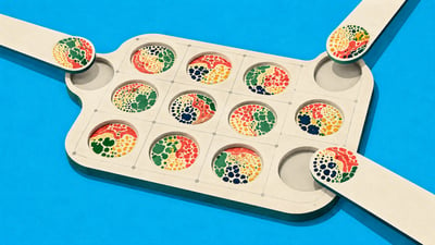
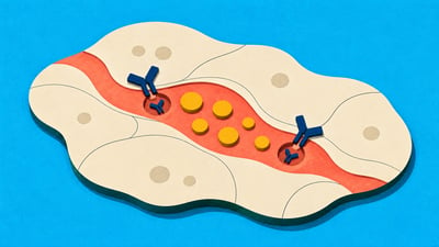
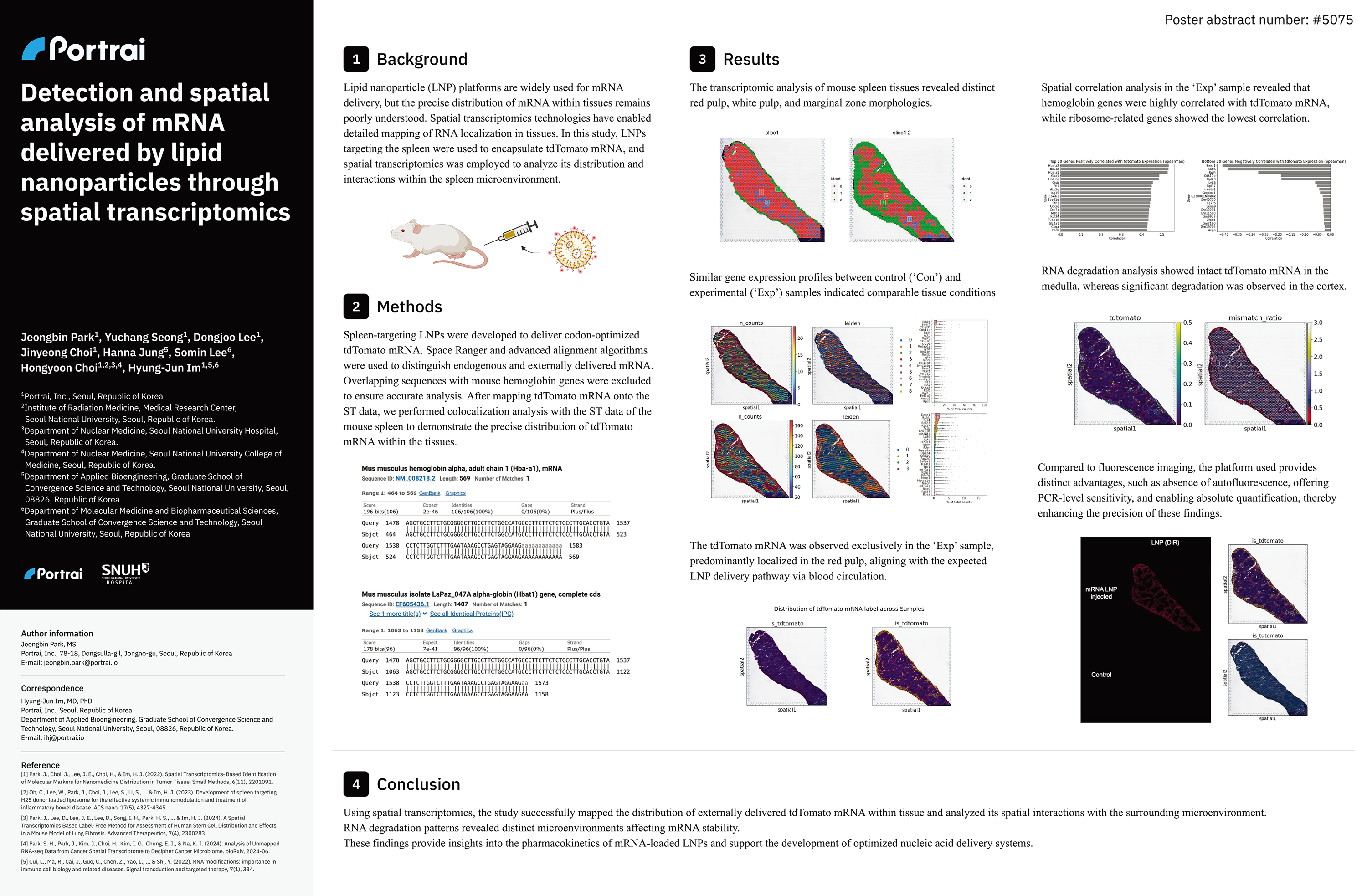
Background: Lipid nanoparticle (LNP) platforms are widely used for mRNA delivery, but the precise distribution of mRNA within tissues remains poorly understood. Spatial transcriptomics technologies have enabled detailed mapping of RNA localization in tissues. In this study, LNPs targeting the spleen were used to encapsulate tdTomato mRNA, and spatial transcriptomics was employed to analyze its distribution and interactions within the spleen microenvironment.
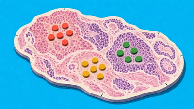
Methods: Spleen-targeting LNPs were developed to deliver codon-optimized tdTomato mRNA. Space Ranger and advanced alignment algorithms were used to distinguish endogenous and externally delivered mRNA. Overlapping sequences with mouse hemoglobin genes were excluded to ensure accurate analysis. After mapping tdTomato mRNA onto the ST data, we performed colocalization analysis with the ST data of the mouse spleen to demonstrate the precise distribution of tdTomato mRNA within the tissues.
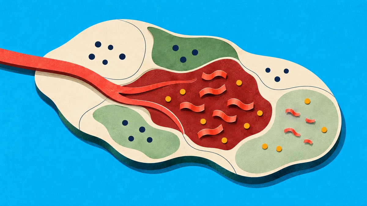
Results: The transcriptomic analysis of mouse spleen tissues revealed distinct red pulp, white pulp, and marginal zone morphologies. Similar gene expression profiles between control (‘Con’) and experimental (‘Exp’) samples indicated comparable tissue conditions. tdTomato mRNA was observed exclusively in the ‘Exp’ sample, predominantly localized in the red pulp, aligning with the expected LNP delivery pathway via blood circulation. Spatial correlation analysis in the ‘Exp’ sample revealed that hemoglobin genes were highly correlated with tdTomato mRNA, while ribosome-related genes showed the lowest correlation. RNA degradation analysis demonstrated intact tdTomato mRNA in the medulla, whereas significant degradation was observed in the cortex. Compared to fluorescence imaging, the platform used provides distinct advantages, such as absence of autofluorescence, offering PCR-level sensitivity, and enabling absolute quantification, thereby enhancing the precision of these findings.
Conclusion: Using spatial transcriptomics, the study successfully mapped the distribution of externally delivered tdTomato mRNA within tissue and analyzed its spatial interactions with the surrounding microenvironment. RNA degradation patterns revealed distinct microenvironments affecting mRNA stability. These findings provide insights into the pharmacokinetics of mRNA-loaded LNPs and support the development of optimized nucleic acid delivery systems.
Authors
Jeongbin Park, Yuchang Seong, Dongjoo Lee, Jinyeong Choi, Hanna Jung, Somin Lee, Hongyoon Choi, Hyungjun Im