One patient is a dataset
Single-Patient Target Discovery with PortraiTARGET
Portrai is building PortraiTARGET, a platform designed to identify the best targets for cancer drugs by analyzing large-scale, human-derived biological data.
Our belief is straightforward. The best cancer drugs should begin with human data. As biological datasets continue to grow in both size and resolution, we see an opportunity to rethink how drugs are discovered and developed. Instead of relying primarily on preclinical models, researchers can first test ideas in silico using comprehensive human molecular data, and then move the most promising candidates into experimental validation and clinical development.
To make this possible, we built PortraiATLAS, a growing collection of tens of thousands of human tissue samples and millions of individual cells. This resource captures the molecular landscape of human cancers at an unprecedented scale and serves as the foundation of PortraiTARGET.
Our long-term vision is to enable human data-driven simulation before the first patient enters a clinical trial, helping researchers identify better drug targets, reduce uncertainty, and accelerate the development of more effective cancer therapies.
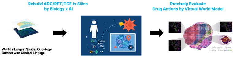
Beyond drug discovery, PortraiTARGET can also support treatment decisions for individual patients.
A great example is the case of Sid, whose cancer journey is shared publicly. Sid was diagnosed with osteosarcoma, a rare cancer of the bone. These data include various types of biomedical data, from single-cell RNA-seq to whole-body imaging. Among them, spatial transcriptomics provides a holistic molecular map that is particularly useful for PortraiTARGET. Two tumor cores were placed on glass slides and analyzed using two different spatial profiling technologies. There was no large patient cohort, no matched controls, and no similar cases to compare against.
Despite its size, this dataset represents exactly the kind of challenge where a human data-driven approach can make a difference. Instead of asking what works on average across thousands of patients, we can ask a much more personal question: what drug is most likely to work for this specific patient?
This is particularly relevant for antibody-drug conjugates (ADCs) and radiopharmaceutical therapies (RPTs). Both treatment modalities depend on finding molecules that are present on the surface of cancer cells but absent, or sufficiently limited, in healthy tissues. These surface molecules serve as entry points, allowing highly potent drugs or radioactive isotopes to be delivered to tumors while minimizing damage to normal cells.
The central question is simple but critical. Is there a molecule on the surface of this patient's cancer cells that a drug can target without affecting healthy tissue?
Using PortraiTARGET, we ranked data-driven potential targets across multiple biological and therapeutic criteria. We then compared these candidates with targets that are already being pursued by pharmaceutical companies and simulated how much therapeutic radiation each target could deliver to this patient's tumor.
The results were encouraging. A few well-established targets performed well, many widely studied targets did not, and several overlooked candidates showed even stronger potential. This illustrates how human molecular data can uncover opportunities that might be missed by conventional approaches and, ultimately, support more personalized cancer treatment.
What we had to work with
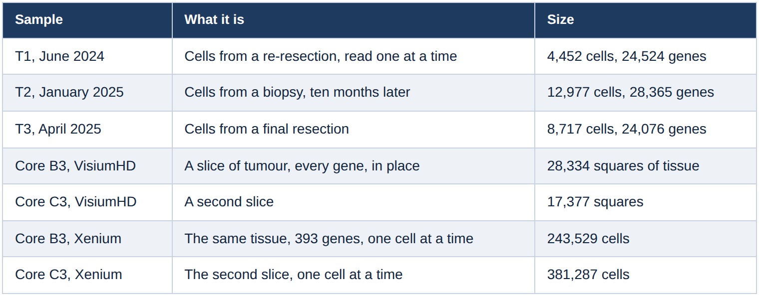
Two things about that table are worth pausing on.
The first three rows are time. Cancer changes under treatment. A target that is present in June and gone by January is a target that will fail in the clinic, and you cannot see that in a single biopsy.
The last four rows are place. A tumor is not a bag of identical cells. It is a structure, with dense regions and sparse ones, with blood vessels and immune cells and scaffolding woven through it. A drug has to physically reach the cancer cells, which means it matters not just whether a target is present but where. The two cores were read twice on purpose: Visium HD reads every gene but pools four to eight cells into each square, while Xenium reads only 393 genes but reads each cell separately. Neither alone answers the question. Together they do.
What a target actually is
A cell is wrapped in a membrane, and studded through that membrane are proteins. Some poke out into the space around the cell. Those are the only proteins a large drug can reach, because antibodies are far too big to get inside a living cell.
That gives you the shape of modern targeted cancer therapy. You can take an antibody that sticks to one specific protein and attach a chemical poison, in which case the result is called an antibody-drug conjugate. For a radioligand, a targeting molecule carries a radioactive isotope to the target. The targeting molecule finds the protein; the payload does the damage.
The whole design rests on one assumption: that the protein is on the cancer and not on anything else. If it is also on your bone marrow, the drug can damage your bone marrow.
So a good target has to clear several bars.
Is it absent from healthy tissue? We answer this by comparing the patient's cancer cells against healthy cells drawn from 28 different tissues of the human body.
Is it on most of the cancer cells, or only some? A drug that reaches 20% of a tumor leaves 80% behind to regrow. This turns out to be the axis that separates the candidates most sharply.
Is it spread through the tissue, or clumped in a corner? Radiation travels a couple of hundred micrometres from where it lands, so a target that sits in scattered islands irradiates the gaps between them and not much else.
Two names to keep in mind
Throughout the series we compare everything against two reference points.
FAP is fibroblast activation protein. It is not on cancer cells at all in most tumors. It is on the fibroblasts that cancer recruits to build its scaffolding, and the idea behind targeting it is that you attack the support structure instead of the tumor. Osteosarcoma, however, originates from mesenchymal cells and can show cancer-cell expression of FAP. Several radioligands against FAP are in clinical trials. Sid also underwent FAP-targeting radiopharmaceutical treatment.
B7-H3, also called CD276, is a protein that appears across many solid tumours including osteosarcoma. There are antibody-drug conjugates and radioligands in clinical trials. If any well-known target should work in this patient, it is this one.
A note on how the work was done
Some of the figures were not made by us. They were made by an AI agent (PortrAIgent) that lives inside this project, was handed the patient's data, proposed its own simulation parameters, waited for a human to approve them, ran the simulation and saved the result. Its working notes are still in the project database. This paper also includes that, because the interesting claim is not that a model can run a script. It is that the reasoning is written down and can be checked.
Mapping a patient in single cells
Before we can ask what is on the cancer cells, we have to know which cells are the cancer. That sounds trivial. It was the hardest part of this study.
The result first
The three biopsies contain 26,146 cells. After harmonising them we can place each one and say what it is.
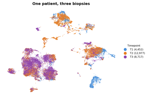
Each point is a cell. Cells that behave similarly land near each other. The three colors are the three biopsies, spread over ten months.
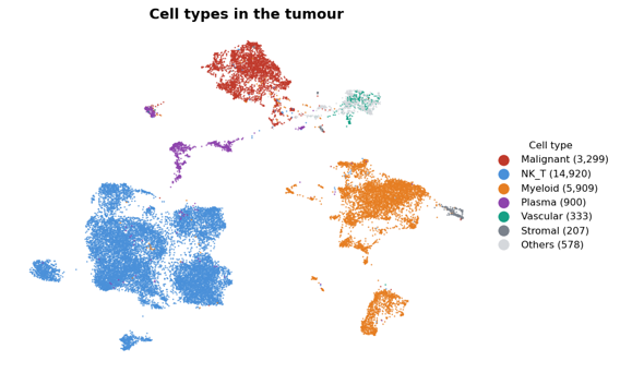
The same map, colored by what each cell turned out to be, separates malignant and non-malignant populations. The cancer cells form a compact island; the remaining populations include T and natural killer cells, myeloid cells, plasma cells, vascular cells, stromal cells and others.
3,299 of the 26,146 cells are cancer. That is the population every later measurement is about.
The whole body as a control
A drug target, particularly for ADCs, RPTs and TCEs(T-cell Engagers), has to be absent from healthy tissue. Absent, or close enough that the dose that kills the tumor does not kill the patient.
To test that for one patient you need a picture of the healthy human body at the same resolution as your tumor. That picture exists. It is called Tabula Sapiens, and the version we used (subsampled for easier analysis) contains 113,621 individual cells drawn from 28 tissues: bone marrow, blood, heart, kidney, liver, lung, skin, both intestines, salivary gland, eye, and eighteen more.
We put the patient's 26,146 cells into the same space as those 113,621 healthy cells and asked what stands out.
The result first
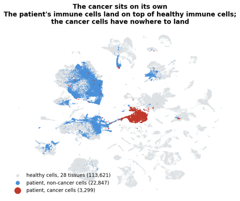
Grey is a healthy human body. Blue is the patient's own immune and vascular cells. Red is the patient's cancer.
The blue lands on top of the grey. The patient's T cells look like healthy T cells, because they are healthy T cells. That overlap is the control experiment: it shows the integration is working, that we have not simply drawn a circle around one sample.
The red does not land on anything.
The funnel
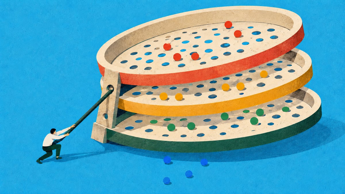
Starting from 8,000 genes, six filters in sequence.
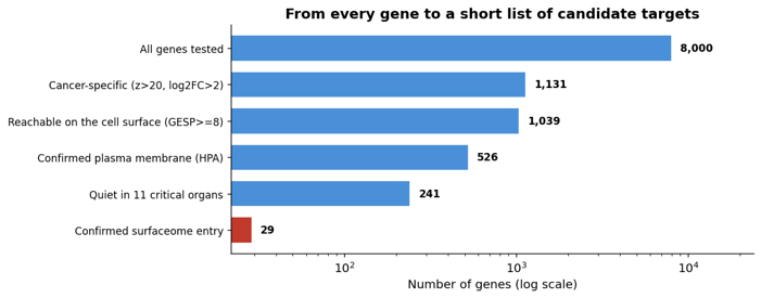
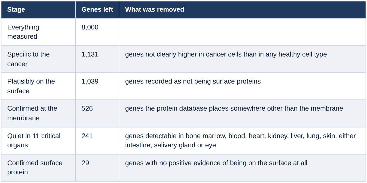
One of those stages deserves explanation.
Quiet in 11 critical organs. The organ list is not arbitrary. These are the tissues that kill you first if a drug lands there. Bone marrow leads the list, and for this patient it matters twice: it is the dose-limiting organ for any radioactive drug. A gene is treated as present in an organ if its average level clears a threshold and more than 5% of that organ's cells carry it. Both conditions. One bright cell in a liver sample is not liver expression. This stage removed more than half of what reached it.
The standard surface filters are written to exclude: they drop a gene if a database records it as not being a surface protein. Of the 241 genes that reached this stage, 212 had no positive surface evidence. Requiring positive evidence takes 241 to 29.
What survived
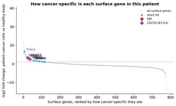
Every surface gene, ranked by how specific it is to the cancer. The blue points are the 29 that cleared every filter. The two diamonds are our reference targets.
The list: ITGA10, ITGA11, GPR158, GPC1, SEMA7A, S1PR3, ADGRA2, NPFFR2, GRM7, NPR2, TBXA2R, GPR173, ASIC1, GABRA3, CDH8, CDH18, SLC24A2, SLCO1A2, SLCO6A1, OR56A1, and nine members of the clustered protocadherin family.
Two of those are worth naming now.
ITGA10 is integrin alpha-10, a collagen receptor found on mesenchymal cells. It is 13-fold higher in this patient's cancer than in the healthy body, and it has almost no signal in any of the 11 critical organs. Looking it up afterwards, there is a published body of work arguing that integrin alpha-10 drives sarcoma growth and is a therapeutic target with narrow normal-tissue distribution, and binders against it have been made and tested in animals. As far as we can find, nothing against it has reached a clinical trial. So this is a preclinical target that somebody else independently arrived at, which our pipeline surfaced from one patient's data without being told to look for it. That is the method doing its job, and it is not the same as a validated drug.
GPC1 is glypican-1, a surface proteoglycan. Antibody-drug conjugates against it have shown activity in animal models of pancreatic, oesophageal and lung cancer. Also preclinical, as far as we can tell.
Where the well known targets land
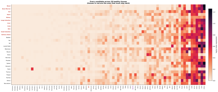
The safety measure is a single number per gene: the average level across the 11 critical organs, on a log scale. Lower is safer.
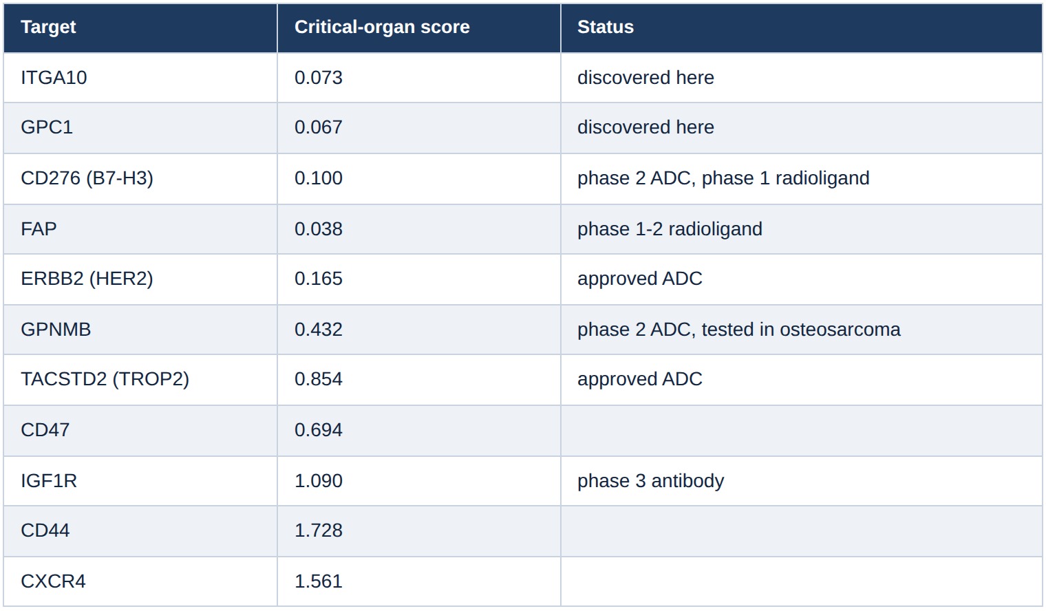
FAP and B7-H3 are both relatively favorable by this measure. TROP2, the target of approved drugs, scores about 22 times higher than FAP here, largely because of broader normal-tissue expression, including eye and salivary gland. That is not news to the people who develop TROP2 drugs, who manage normal-tissue liabilities through molecule design and dosing, but it tells you the number is measuring something real.
The bottom of that table is the interesting part. CD44, CXCR4, and IGF1R are all things people have proposed as targets, and all three are broadly expressed in the healthy body. Whatever their merit elsewhere, a whole-body reference rules them out for this patient before any tumor measurement is taken.
What this stage does not tell you
Safety is a veto, not a recommendation. Every one of the surviving targets passed because it is absent, or sufficiently limited, in healthy tissue, and a gene can be absent from healthy tissue and also nearly absent from the tumour.
Three of the 29 are on fewer than 2% of the patient's cancer cells. They cleared every filter in this post and they are useless.
Sources for the claims about other people's work
Anti-glypican-1 antibody drug conjugate against pancreatic cancer
Glypican-1-targeted antibody drug conjugate in pancreatic and oesophageal cancer
This case study is for research and exploratory purposes only. The results have not been clinically validated and should not be used to guide patient care.