doi: https://aacrjournals.org/cancerres/article/85/8_Supplement_1/2443/760927
Poster
Abstract
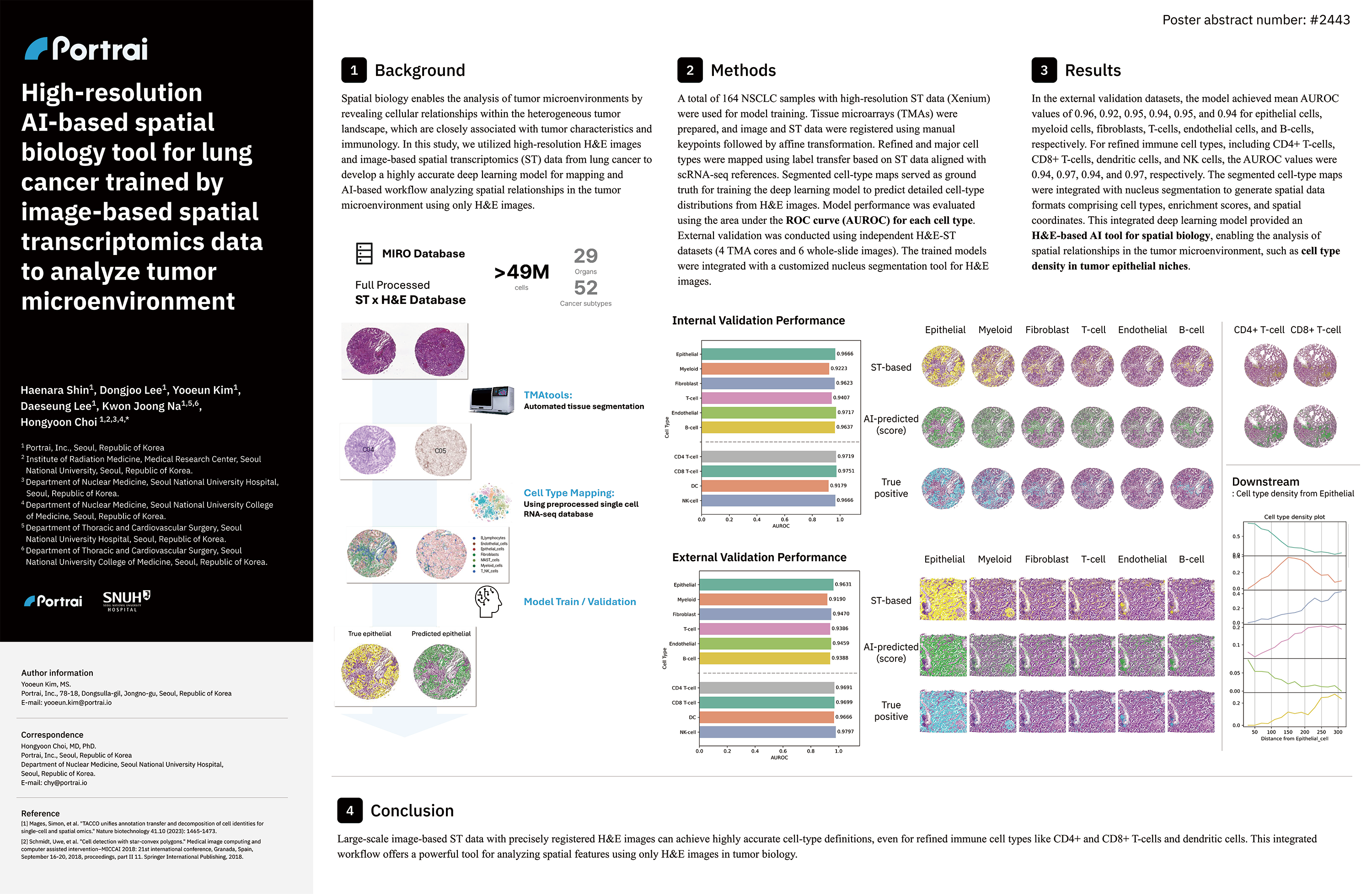
Background: Spatial biology enables the analysis of tumor microenvironments by revealing cellular relationships within the heterogeneous tumor landscape, which are closely associated with tumor characteristics and immunology. In this study, we utilized high-resolution H&E images and image-based spatial transcriptomics (ST) data from lung cancer to develop a highly accurate deep learning model for mapping and AI-based workflow analyzing spatial relationships in the tumor microenvironment using only H&E images.
Methods: A total of 164 NSCLC samples with high-resolution ST data (Xenium) were used for model training. Tissue microarrays (TMAs) were prepared, and image and ST data were registered using manual keypoints followed by affine transformation. Refined and major cell types were mapped using label transfer based on ST data aligned with scRNA-seq references. Segmented cell-type maps served as ground truth for training the deep learning model to predict detailed cell-type distributions from H&E images. Model performance was evaluated using the area under the ROC curve (AUROC) for each cell type. External validation was conducted using independent H&E-ST datasets (6 TMA cores and 4 whole-slide images). The trained models were integrated with a customized StarDist-based nucleus segmentation tool for H&E images.
Results: In the external validation datasets, the model achieved mean AUROC values of 0.96, 0.92, 0.95, 0.94, 0.95, and 0.94 for epithelial cells, myeloid cells, fibroblasts, T-cells, endothelial cells, and B-cells, respectively. For refined immune cell types, including CD4+ T-cells, CD8+ T-cells, dendritic cells, and NK cells, the AUROC values were 0.94, 0.97, 0.94, and 0.97, respectively. The segmented cell-type maps were integrated with nucleus segmentation to generate spatial data formats comprising cell types, enrichment scores, and spatial coordinates. This integrated deep learning model provided an H&E-based AI tool for spatial biology, enabling the analysis of spatial relationships in the tumor microenvironment, such as cell type density in tumor epithelial niches.
Conclusion: Large-scale image-based ST data with precisely registered H&E images can achieve highly accurate cell-type definitions, even for refined immune cell types like CD4+ and CD8+ T-cells and dendritic cells. This integrated workflow offers a powerful tool for analyzing spatial features using only H&E images in tumor biology.
Authors
Haenara Shin, Dongjoo Lee, Yooeun Kim, Daeseung Lee, Kwon Joong Na, Hongyoon Choi.