How Far Can We Move from “Hypothesis-Driven DDS” to “Spatially Precise DDS”?
Over the past two decades, tumor drug delivery systems (DDS), particularly nanomedicines, have advanced rapidly based on the expectation that better materials would enable drugs to reach tumors more effectively. Yet a persistent clinical challenge remains: delivery performance and therapeutic response vary even when the same platform and drug are used to treat the same cancer type.
Tumors are too complex for this inconsistency to be attributed to a single cause, such as the limited effect of EPR (enhanced permeability and retention, the principle underlying the passive accumulation of nanoparticles in tumors) in humans. Instead, a more compelling view is emerging: tumors are spatially compartmentalized ecosystems, and drug delivery is determined by the architecture of those compartments.
In this article, “spatial biology” refers broadly to an approach that combines single-cell analysis, spatial transcriptomics, spatial proteomics, and tissue imaging to read tissues as maps. Its potential to transform DDS research lies in enabling engineering that uses the spatial architecture of tumor tissue as a design input, extending beyond materials engineering focused solely on improving nanoparticles.
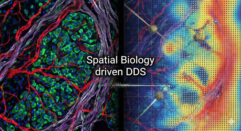
Spatial Biology-Based Drug Delivery System Development (Generated with Gemini Nano Banana)
1) Why Is Delivery So Challenging in Tumors? Barriers Are Spatial Patterns, Not Average Values
From a drug delivery perspective, the tumor microenvironment (TME) is commonly described as a combination of the following barriers:
Abnormal vasculature, including heterogeneous perfusion and permeability and altered pericyte coverage
Increased extracellular matrix (ECM) and fibrosis, or stroma, accompanied by elevated interstitial fluid pressure (IFP)
Immunosuppressive cells, including TAMs, MDSCs, and Tregs, and immune-exclusion structures
What matters, however, is not simply whether these barriers exist, but how they are spatially arranged within the tumor. Even within the same tumor:
Drugs may readily reach perivascular regions
Dense, stroma-rich regions may impede their movement
Hypoxic tumor cores may present distinct delivery conditions
Immune-exclusion boundaries may alter delivery pathways entirely
Drug delivery is therefore a function of both material properties and spatial architecture.
A single metric such as the amount of a drug that enters a tumor (%ID/g) will no longer be sufficient. Where the drug reaches and how uniformly it is distributed, measured through coverage and uniformity, are likely to become increasingly important indicators of performance.
2) Where Conventional DDS Strategies Reach Their Limits: Design Assumptions Are Vulnerable to Spatial Heterogeneity
The major DDS strategies to date can broadly be summarized as follows:
EPR-based passive accumulation
Ligand-based active targeting through receptor binding
TME-responsive release triggered by conditions such as pH, hypoxia, or enzymes
These strategies will remain relevant. From a research perspective, however, their inconsistent reproducibility can often be traced to the failure of spatial assumptions.
Knowing that a target receptor is expressed is insufficient. What matters is where its expression is concentrated, how patchy the distribution is, and whether the receptor is accessible.
Even when vascular permeability is high, dense ECM and elevated IFP may prevent penetration into the tumor interior.
Boundaries created by immune-stromal interactions may function as exclusion zones that block penetration.
Until now, researchers have typically developed a delivery vehicle first and then adjusted it after assessing whether it reaches the tumor. In the future, this sequence may be reversed. Researchers could map the tissue first and design the delivery vehicle around that map.
3) What Spatial Biology Could Enable: Turning Target, Barrier, and Trigger into Design Maps
The most practical contributions of spatial biology to DDS design can be divided into three categories.
(A) Target Map: Not Just “What to Target,” but “Where the Target Is”
As spatial transcriptomic and proteomic maps become more comprehensive, target selection could evolve from choosing receptors based primarily on high average expression to a multi-objective optimization problem incorporating:
Coverage across the entire tumor
Cell type specificity
Spatial continuity or patchiness
Distance from blood vessels and therefore accessibility
This could make it possible to design strategies such as dual- or multi-targeting, in which different targets are combined for different niches, and sequential targeting, in which stromal or immune cells are modulated before tumor cells are targeted. Such strategies could be guided by spatial evidence rather than intuition alone.
(B) Barrier Map: Identifying the Cause of Each Barrier and Selecting an Intervention Point
If spatial data can translate barriers from qualitative descriptions into quantitative metrics, researchers may be able to determine whether poor intratumoral penetration is caused primarily by:
Vascular factors, including perfusion and permeability
ECM or elevated IFP
Immune-exclusion boundaries
This distinction could support a more rational selection of delivery properties, including size, elasticity, surface characteristics, and shape, as well as combination strategies such as stromal modulation, vascular normalization, and immune remodeling.
(C) Trigger Map: Release Becomes a Question of “Where It Turns On”
Stimulus-responsive DDS will remain important, but the criteria for selecting triggers may change.
Triggers such as pH, hypoxia, and protease activity are not distributed uniformly throughout a tumor. As spatial multi-omics advances, triggers could be defined according to spatial conditions:
Strong within the niche where the drug should be activated (ON-site)
Weak in normal tissues or unintended niches (OFF-site)
This could enable more precise control over drug release.
4) Outlook: How Could Spatially Precise DDS Transform Research and Development?
Although these approaches have not yet been standardized, several changes are likely to emerge gradually across research and development.
Change 1) DDS Development Begins with a Map, Not a Formulation
Conventional approach: Platform design → Animal studies → Outcome-based adjustment
Emerging approach: Spatial profiling → Niche and barrier definition → Design hypothesis → Formulation development → Spatial PK/PD validation
Change 2) Performance Metrics Expand from Accumulation to Distribution
Future assessments of intratumoral distribution may combine metrics such as:
Radial distribution based on distance from blood vessels
Exposure within specific niches, such as CAF-rich, immune-rich, and hypoxic regions
Cell type-specific uptake by tumor cells, TAMs, and CAFs
Uniformity metrics, including the coefficient of variation, entropy, and Gini-like indices
Researchers may increasingly seek to establish systematic relationships between these distribution characteristics and therapeutic response through spatial PK/PD-efficacy correlations.
Change 3) Subtype-Specific Delivery Vehicles and Modular Platforms Become Practical Goals
Given the substantial spatial heterogeneity of tumors, a perfect one-size-fits-all solution is unlikely. A more practical roadmap may be a modular DDS in which ligands, release mechanisms, and material properties are combined according to subtypes defined by spatial features, including:
Tumor or immune phenotypes, such as immune-hot and immune-cold tumors
Degree of fibrosis
Hypoxia patterns
5) Challenges Ahead: Research Questions That Must Be Addressed
Several technical and conceptual challenges must be resolved to translate this vision into practice.
Standardization and quantification of spatial data: Maps may appear different depending on the experimental platform or analytical pipeline.
Causal connections between spatial maps and delivery parameters: Models must explain why particular patterns occur rather than merely identify correlations.
Systematic experimental validation: Reproducible protocols are needed to measure and compare spatial PK/PD.
Translational relevance: Researchers must determine how closely the spatial architecture of animal models reflects that of patients and how far findings can be extrapolated.
Conclusion: Can Spatial Biology Translate DDS into the Language of Precision Medicine?
Spatial biology is not simply a way to make DDS more sophisticated. It represents an effort to interpret the reasons for DDS failure through spatial architecture and convert that understanding into actionable design inputs.
Although the field has not yet established fully standardized approaches, continued research could gradually shift DDS development in the following directions:
One-size-fits-all systems → Niche-specific, modular platforms
Accumulation-centered assessment → Distribution-, coverage-, and cell type exposure-centered assessment
Empirical optimization → Spatial data-driven hypothesis and validation loops
If this approach becomes embedded in the development process, the reproducibility challenges of tumor drug delivery may no longer remain variables left to chance. Instead, many of them could become spatially defined and technologically addressable engineering problems.