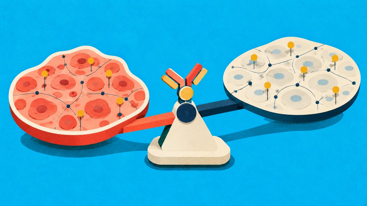
Why Does an Effective Drug Put Patients at Risk?
The cruelest paradox in drug development is that the more effective a drug, the stronger its toxicity can be.
Take ADCs (antibody-drug conjugates) as an example. ADCs are designed to deliver a potent cytotoxic payload to tumors by binding to a target. But what if that target is also expressed in normal tissues? What if the payload reaches not only the tumor but also the heart, lungs, and liver?
This is on-target off-tumor toxicity — the most difficult-to-predict and most fatal problem in ADC development.
In clinical practice, this problem is already a reality:
Cardiotoxicity of HER2-targeting agents: Cardiomyocytes express HER2, where HER2/HER4 heterodimers activated by neuregulin-1 (NRG-1) signaling play a critical role in cardiomyocyte survival and stress resistance. Blocking HER2 disrupts this cardioprotective pathway, leading to increased ROS accumulation and cardiomyocyte apoptosis (Eaton & Timm, Cardio-Oncology, 2023).
Interstitial lung disease (ILD) associated with ADCs: A recent study demonstrated that ADCs are taken up by CD68+ alveolar macrophages in lung tissue via Fcγ receptor-mediated uptake, releasing cytotoxic payload — reported as a major mechanism of ADC-induced ILD (Kumagai et al., Molecular Cancer Therapeutics, 2025). Notably, ILD occurrence does not necessarily correlate with target antigen expression levels in lung tissue; linker and payload characteristics may be more important determinants (Desai et al., Cancers, 2024).
Payload-dependent toxicities: ADCs with MMAF and DM4 payloads show high rates of ocular toxicity (corneal microcystic changes, etc.), while MMAE (tubulin-binding) payloads are associated with peripheral neuropathy. These payload–toxicity associations have been confirmed through meta-analyses and FAERS pharmacovigilance data (Zhu et al., Cancer, 2023; Tang et al., Frontiers in Pharmacology, 2024).
These toxicities often surface only in Phase 1–2 trials, and sometimes lead to trial suspension or market withdrawal. Billions of dollars in investment can evaporate due to a single toxicity issue.
Let us pose the fundamental question: Why can we not predict this toxicity in advance?
The Black Box of Therapeutic Index
In drug development, the Therapeutic Index (TI) is the most fundamental metric. The ratio between the concentration that produces therapeutic effect and the concentration that causes toxicity — the wider this window, the safer the drug.
Yet conventional TI assessment has fundamental limitations:
Tumor side (efficacy): Target expression is quantified via IHC H-score, FISH, etc. → However, as discussed in the March blog, target "quantity" alone cannot reliably predict actual drug delivery and response.
Normal tissue side (toxicity): Toxicity evaluation in preclinical animal models, limited IHC to confirm normal tissue expression → However, normal tissue expression patterns often differ between animals and humans, and IHC does not provide spatial information about which cell types are exposed and to what degree.
As a result, both sides of TI — efficacy and toxicity — are being calculated with spatial context missing. This is the root cause of unexpected toxicities emerging in the clinic.
Spatial Atlas of Normal Tissues: Reading the "Shadow" of the Drug
Spatial biology provides the key to open this black box. The core idea is to perform the same level of spatial analysis on normal tissue as we do on tumors.
By building spatial transcriptomics data on normal heart, lung, liver, and kidney tissue, we can answer the following questions:
1. Which Cells Are Exposed to the Payload?
Just as an ADC bound to its target releases payload in the tumor, payload can also be released from target-expressing cells in normal tissues. However, the key point is that knowing "the target is expressed" alone is insufficient to predict toxicity.
In spatial transcriptomics data, we can identify the exact cell type and spatial location of target-expressing cells within normal tissues. For example:
In the heart, HER2 is primarily expressed in ventricular cardiomyocytes → direct payload exposure to these cells poses cardiotoxicity risk. Disruption of the HER2/HER4-NRG1 cardioprotective pathway is the key mechanism (Eaton & Timm, 2023)
In the lung, CD68+ alveolar macrophages take up ADCs via Fcγ receptor and release payload → a major cellular mechanism of ILD (Kumagai et al., 2025). TROP2 expression has also been confirmed in alveolar epithelial cells (AT1/AT2), though a direct causal link to ILD is still under investigation
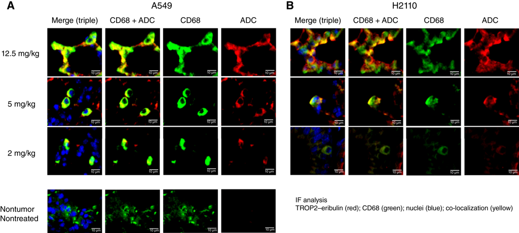
Figure 1. Spatial distribution of ADC and alveolar macrophages in normal lung tissue. Immunofluorescence staining showing dose-dependent accumulation of a TROP2-targeting ADC (red) in CD68+ alveolar macrophages (green) within lung tissue. Blue indicates nuclei (DAPI). ADC uptake by macrophages occurred regardless of target antigen expression levels in the lung, suggesting a key spatial mechanism underlying on-target off-tumor toxicity. (Adapted from Kumagai et al., Molecular Cancer Therapeutics, 2025; Fig. 3)
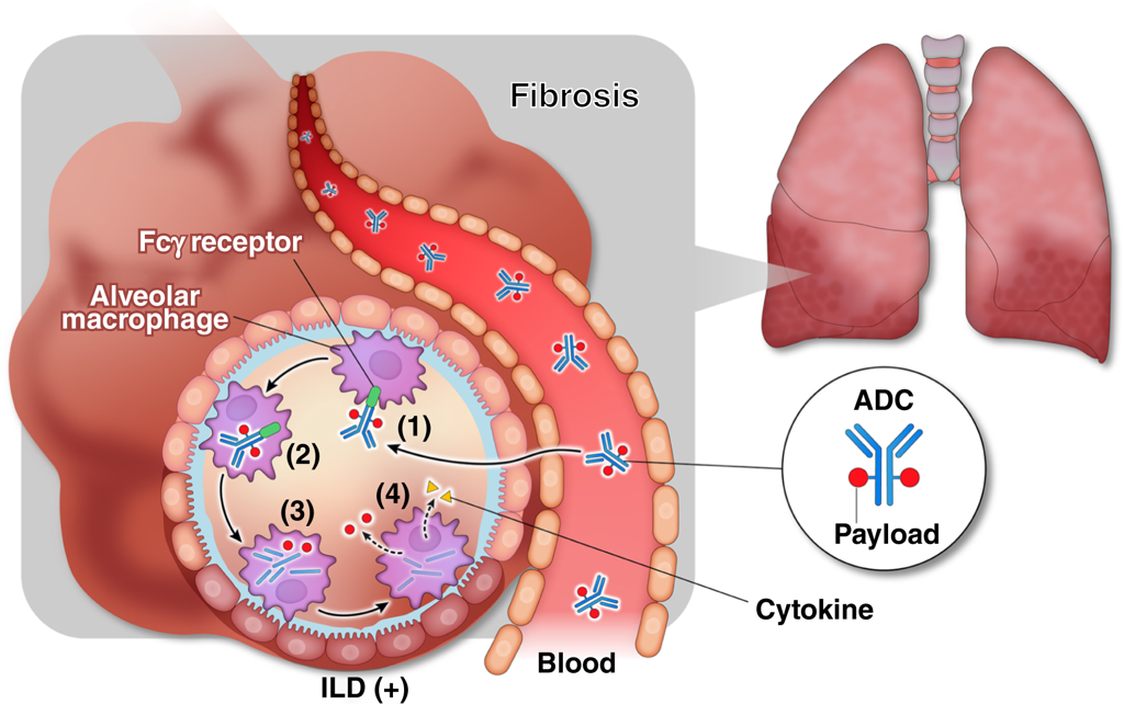
Figure 2. Proposed mechanism of ADC-induced interstitial lung disease (ILD). (1) ADC binds to Fcγ receptors on the surface of alveolar macrophages, (2) is taken up via Fcγ receptor-mediated endocytosis, (3) undergoes intracellular degradation, and (4) releases the cytotoxic payload from macrophages into lung tissue, potentially leading to cytokine-mediated lung injury. This nonspecific pathway occurs independently of target antigen expression, demonstrating that normal tissue toxicity can arise through mechanisms distinct from on-tumor efficacy. (Adapted from Kumagai et al., Molecular Cancer Therapeutics, 2025; Fig. 5)
2. How Far Does the Payload Spread Beyond Target-Positive Cells?
The bystander effect discussed in the March blog is a powerful weapon in tumors, but in normal tissues it becomes a pathway for toxicity spread.
When payload released from a small number of target-expressing normal cells affects surrounding target-negative cells, the actual scope of toxicity can be far broader than what the number of target-expressing cells would predict. By measuring the spatial distance and density between target-positive and surrounding cells in spatial data, the extent of bystander-mediated toxicity can be estimated.
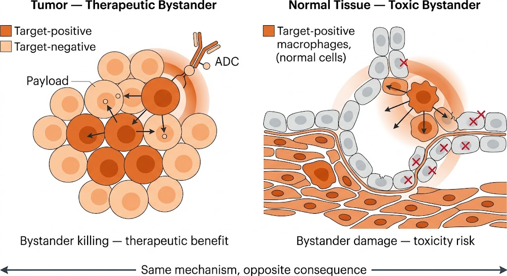
Figure 3. The two faces of the bystander effect: therapeutic benefit in tumors vs. toxicity risk in normal tissues. (Left) In tumor tissue, payload released from an ADC bound to a target-positive cell (dark orange) diffuses to surrounding target-negative cells (light orange), inducing bystander killing — a therapeutic advantage. (Right) In normal tissue, payload released from a small number of target-positive cells (e.g., macrophages, cardiomyocytes) diffuses to surrounding healthy cells, causing tissue damage — a toxicity risk. Payload concentration decreases with distance from the release point (gradient), and the same bystander mechanism determines efficacy coverage in tumors and toxicity exposure in normal tissues. (Created by AI)
3. How Does Vascular Architecture Alter Drug Exposure?
Normal tissue vascular density and structure differ fundamentally from tumors. Unlike the abnormal vasculature (leaky vasculature) of tumors, normal tissue vessels generally have normal permeability.
However, because vascular density and arrangement differ by organ, the same dose of ADC results in different drug exposure levels across normal organs. The heart is a highly vascularized organ, meaning ADC exposure may be high — this forms the anatomical basis for cardiotoxicity.
By comprehensively analyzing vascular density, target expression location, and spatial relationships of major cell types in each normal organ using spatial transcriptomics data, organ-specific toxicity risk can be quantitatively estimated.
Examining the Therapeutic Window Through a Spatial Lens
When these three analyses — cell type-specific target expression, bystander diffusion range, and vascular–drug exposure architecture — are performed simultaneously on both tumor and normal tissue, we can evaluate the therapeutic window with far greater spatial precision.
The conventional therapeutic index is defined as:
Conventional approach = Toxic dose / Therapeutic dose (based on systemic pharmacokinetics)
With spatial data, we can examine this at the tissue level with much finer granularity:
Comparing intratumoral payload reach (efficacy coverage) vs. normal tissue payload exposure (toxicity exposure) within the same spatial framework
In this approach:
Efficacy coverage: The proportion of cells within the tumor where payload reaches therapeutic concentration (above IC50). Determined by vessel-to-target distance, linker–enzyme matching, and bystander range (March blog)
Toxicity exposure: The proportion of cells in normal tissue where payload exceeds the toxicity threshold. Determined by target-expressing cell types, bystander diffusion, and vascular density (this blog)
Comparing both values within the same spatial framework allows us to answer the most fundamental question in ADC design: "Can this drug kill the tumor sufficiently while preserving normal tissue sufficiently?"
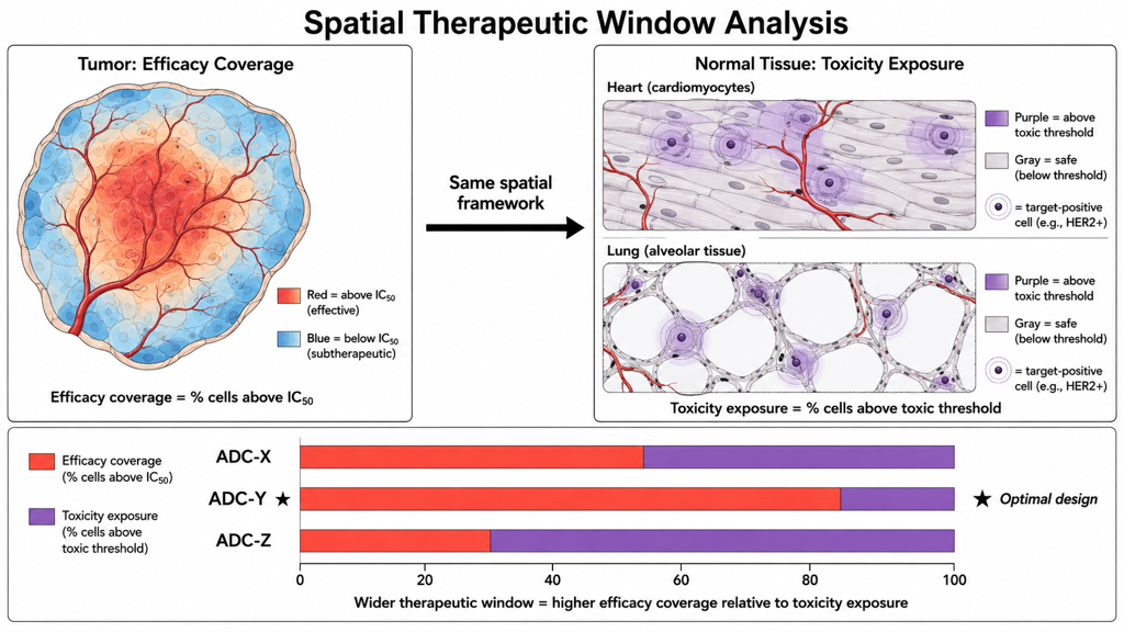
Figure 4. Spatial therapeutic window analysis framework. (Left) Efficacy coverage in tumor tissue — regions where payload reaches therapeutic concentration above IC50 (red/warm colors) and subtherapeutic regions (blue/cool colors). Payload delivery varies with distance from vascular structures (red lines). (Right) Toxicity exposure in normal tissues (heart, lung) — regions where payload exceeds the toxicity threshold around target-positive cells (purple) and safe regions (gray). (Bottom) Comparison of efficacy coverage (red) vs. toxicity exposure (purple) across different ADC designs (ADC-X, ADC-Y, ADC-Z). The design that maintains efficacy while minimizing toxicity secures the widest therapeutic window. (Created by AI)
Previewing Toxicity In Silico
The most powerful aspect of this spatial analysis approach is the ability to simulate toxicity profiles in silico before actual clinical trials.
Using spatial transcriptomics data from patient tumor tissue and normal tissue, and inputting the physicochemical parameters of the ADC (binding affinity, linker type, payload permeability, DAR, etc.):
Tumor side: TME-PK™ modeling (covered in the March blog) calculates intratumoral payload distribution and efficacy coverage
Normal tissue side: The same modeling framework is applied to normal tissue to calculate organ-specific toxicity exposure
Therapeutic window evaluation: Both results are integrated to assess the spatial efficacy-toxicity balance of ADC candidates
This approach can be directly applied to ADC design optimization:
How does changing the linker alter payload release in normal lung tissue?
Does tuning binding affinity (Kd) reduce drug exposure in the heart?
Between payloads with strong vs. weak bystander effects, which has a wider toxicity margin in normal tissue?
If we can answer these questions before animal studies or clinical trials, we could meaningfully reduce ADC development failure rates.
Conclusion: The Era of Seeing Both Sides of the Therapeutic Window
Until now, ADC development has focused on efficacy while managing toxicity relatively reactively. All efforts were directed at tumor response, while normal tissue toxicity was handled with a "respond when observed" approach in clinical trials.
Spatial biology can change this paradigm. By analyzing tumor and normal tissue in the same spatial language, it is becoming possible to examine both efficacy and toxicity simultaneously, enabling more precise therapeutic window evaluation before entering the clinic.
As we confirmed at AACR in April, spatial biology has become the language for reading drug response and resistance. This month, I discussed how to read the other side of that language — the shadow left by the drug: toxicity.
Only when we look not just at the "light" of the drug but also at its "shadow" can we take one step closer to true precision medicine.
References
Eaton H, Timm KN. Mechanisms of trastuzumab induced cardiotoxicity -- is exercise a potential treatment? Cardio-Oncology. 2023;9:22. DOI: 10.1186/s40959-023-00172-3
Kumagai K, et al. Potential Mechanisms of Interstitial Lung Disease Induced by Antibody-Drug Conjugates Based on Quantitative Analysis of Drug Distribution. Molecular Cancer Therapeutics. 2025;24(2):242-250. DOI: 10.1158/1535-7163.MCT-24-0267
Desai A, Subbiah V, Roy-Chowdhuri S, et al. Association of Antibody-Drug Conjugate Target Expression and Interstitial Lung Disease in Non-Small-Cell Lung Cancer. Cancers. 2024;16(22):3753. DOI: 10.3390/cancers16223753
Zhu Y, Liu K, Wang K, Zhu H. Treatment-related adverse events of antibody-drug conjugates in clinical trials: A systematic review and meta-analysis. Cancer. 2023;129(2):283-295. DOI: 10.1002/cncr.34507
Tang X, et al. A pharmacovigilance study on antibody-drug conjugate-related neurotoxicity based on the FDA adverse event reporting system. Frontiers in Pharmacology. 2024;15:1362484. DOI: 10.3389/fphar.2024.1362484
Khera E, Dong S, Huang H, et al. Cellular-Resolution Imaging of Bystander Payload Tissue Penetration from Antibody-Drug Conjugates. Molecular Cancer Therapeutics. 2022;21(2):310-321. DOI: 10.1158/1535-7163.MCT-21-0580
Weddell J, Chiney MS, Bhatnagar S, et al. Mechanistic Modeling of Intra-Tumor Spatial Distribution of Antibody-Drug Conjugates. Clinical and Translational Science. 2021;14(1):395-404. DOI: 10.1111/cts.12892