Portrai’s Next Step in Precision DDS
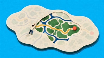
When designing precision drug delivery systems (DDS), the key question is no longer simply, “Does the drug reach the tumor?” We must also examine where within the tumor it reaches and which cells actually take it up. Conventional biodistribution analysis reveals drug accumulation at the organ level, but has limitations in explaining cell-level selectivity within the tumor microenvironment.
Why Spatial Context Matters
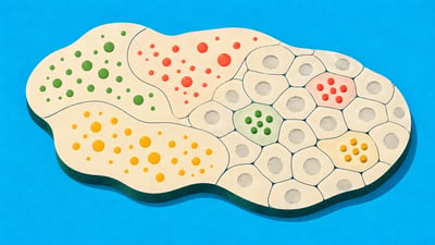
Drug imaging shows where a drug is distributed within tissue, while Spatial Transcriptomics (ST) reveals gene expression and cellular composition at those locations. Integrating the two allows us to interpret both “where the drug goes” and “which biological features are associated with its distribution.”
This concept was presented in a 2022 paper in Small Methods. By integrating fluorescence-based images of nanomedicine distribution with ST data, the study demonstrated that molecular markers related to hypoxia, glycolysis, and apoptosis could be identified in association with nanomedicine accumulation in 4T1 tumor tissue.
From Drug Signals to Mechanistic Insight
Portrai’s approach goes beyond simply overlaying images and omics data. The key is to align drug signals and ST data within a shared spatial coordinate system, then progressively interpret cellular composition and molecular signatures. This enables us to identify which cell populations occupy regions of high drug accumulation and which receptor axes or metabolic programs are associated with those regions.
Core Algorithms Behind the Integrated Analysis Pipeline
The integrated analysis uses several of Portrai’s core algorithms.
SPADE links tissue image features with spatial gene expression, enabling the joint interpretation of drug fluorescence patterns and transcriptomic features.
CellDART estimates the cellular composition of each spatial spot using single-cell RNA sequencing (scRNA-seq) reference data.
IAMSAM uses the Segment Anything Model to extract regions of interest (ROIs) with high drug signals and analyze their molecular signatures.
Together, these algorithms move beyond visualizing drug distribution to interpreting uptake-associated biology at the tissue level.
A Case Study in Glycosylation-Directed Targeting
This framework was also applied in a study of a precision-glycosylated clickable albumin nanoplatform (CAN-DGIT), recently reported in Theranostics (see figure). The study combined PET with ST-based analyses using SPADE, CellDART, and IAMSAM to show how glycosylation type changes pharmacokinetics and cellular selectivity.
Specifically, mannosylated albumin (Man-Alb) showed enrichment in clusters rich in extracellular matrix (ECM) and tumor-associated macrophages (TAMs), as well as in Mrc1-high regions. In contrast, galactosylated albumin (Gal-Alb) and glucosylated albumin [Glc(6)-Alb] showed uptake in glycolytic or hypoxic tumor clusters and Slc2a1-high regions.
Based on these findings, the authors interpreted precisely controlled glycosylation as a means of enabling programmable biodistribution and cell-type targeting within the tumor microenvironment (TME).
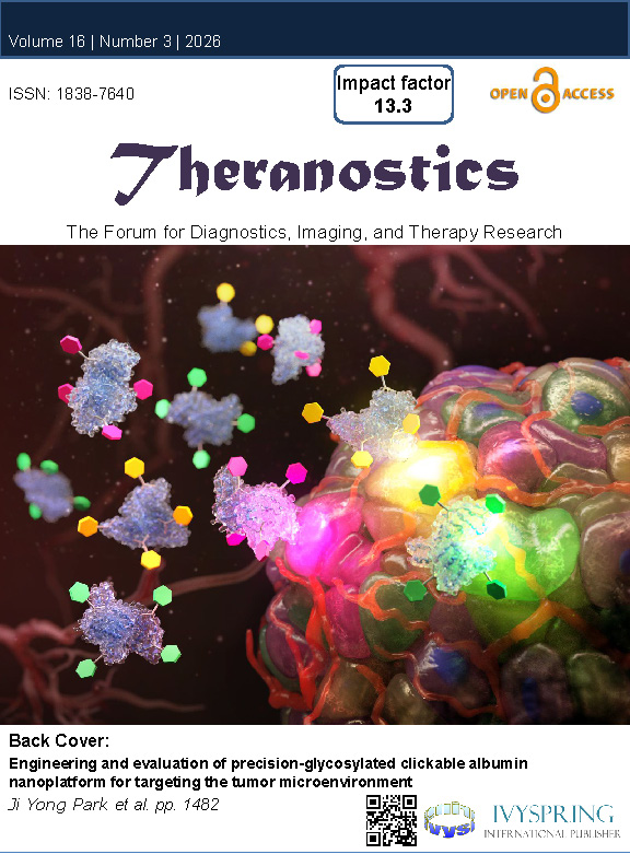
Cover of the corresponding issue of Theranostics
What This Changes for DDS Development
This type of analysis changes the criteria for evaluating DDS. What matters is not simply the amount of drug accumulated in a tumor, but also which niches it reaches, which cells receive the payload, and which molecular programs are associated with that selectivity.
Integrating ST with drug imaging supports more precise comparisons of drug candidates, optimization of ligand design, and prioritization of follow-up validation.
The Next Step in Precision Drug Delivery
Portrai views Spatial Transcriptomics not simply as an omics readout, but as a functional spatial layer for interpreting drug delivery. This approach could extend beyond glycoengineered nanoparticles to ligand-directed DDS, antibody–drug conjugates (ADCs), radioligand delivery, and immune-cell-targeted therapeutics.
The next step in precision DDS is to move beyond drugs that accumulate in large quantities toward designing and validating drugs that reach the right cells and niches.
References
Park J, Choi J, Lee JE, Choi H, Im HJ. Spatial Transcriptomics-Based Identification of Molecular Markers for Nanomedicine Distribution in Tumor Tissue. Small Methods. 2022;6(11):e2201091. doi:10.1002/smtd.202201091.
Bae S, et al. Discovery of molecular features underlying the morphological landscape by integrating spatial transcriptomic data with deep features of tissue images. Nucleic Acids Research. 2021. SPADE.
Bae S, et al. CellDART: cell type inference by domain adaptation of single-cell and spatial transcriptomic data. Nucleic Acids Research. 2022;50(10):e57.
Lee D, Park J, Cook S, et al. IAMSAM: image-based analysis of molecular signatures using the Segment Anything Model. Genome Biology. 2024;25(1):290.
Park JY, et al. Engineering and evaluation of precision-glycosylated clickable albumin nanoplatform for targeting the tumor microenvironment. Theranostics. 2026.