Where Does the Uncertainty for Patients and Physicians Come From?
For physicians treating cancer in clinical practice, the most difficult moment is not necessarily when surgery technically fails. It may be when cancer recurs despite treatment following standard guidelines, with the disease progressing differently from what was predicted.
Consider two patients, A and B, who both have early-stage, or Stage I, lung cancer. According to the TNM staging system, their cancers are identical. Both tumors are smaller than 3 cm, and neither patient has lymph node metastasis. Following current standards of care, including the NCCN and ESMO Guidelines, both patients undergo surgery and are monitored every six months without additional adjuvant chemotherapy.
Patient A remains recurrence-free for five years, is declared cured, and returns to everyday life. Patient B, however, experiences a recurrence after 18 months and must begin systemic chemotherapy.
In hindsight, Patient B may have needed additional adjuvant therapy immediately after surgery or more frequent surveillance. Yet the current TNM staging system provided no basis for making that distinction.
Bridging this gap requires more than simply collecting additional data. Physicians need interpretable data that allows them to make treatment decisions with confidence. This is why I want to discuss spatial biology.
The Limits of the Map: Anatomy Does Not Fully Represent Biology
For decades, the TNM staging system, which stands for Tumor, Node, and Metastasis, has served as the primary compass for cancer treatment. It classifies cancer from Stage I to Stage IV according to the size or extent of the primary tumor (T), whether it has spread to nearby lymph nodes (N), and whether it has metastasized to other organs (M). It is an excellent system for describing the geographical extent of cancer.
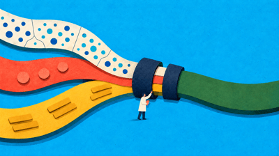
However, this map does not reveal one of cancer’s most important characteristics: its underlying biology.
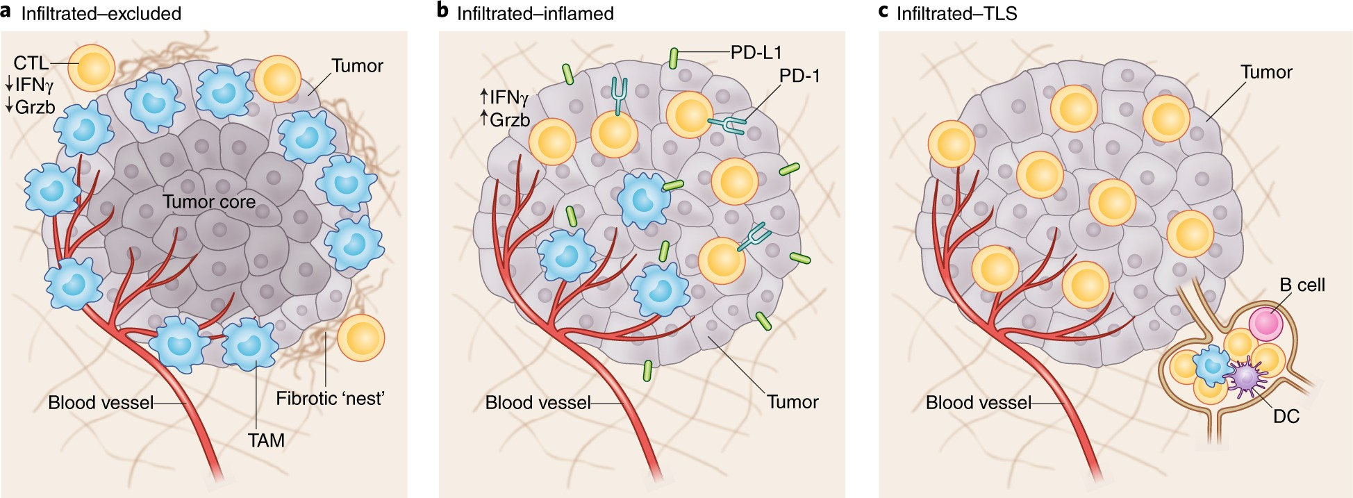
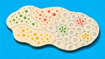
Proposed classification of the Tumor Immune Microenvironment (TIME) (Nature Medicine, 2018)
As the figure illustrates, even Stage I cancers that appear identical can differ dramatically internally. Some remain indolent, while others behave aggressively. I believe Patient B’s cancer recurred not because of its size, but because the way the cancer cells interacted with their surroundings, namely the tumor microenvironment (TME), was fundamentally different from that of Patient A.
We must now look beyond tumor size to examine cellular composition and spatial context.
Opening the Black Box: Reading Spatial Context
To investigate this mystery, clinical researchers began examining pathology slides, including H&E-stained tissue sections, in much greater detail. They counted the immune cells, fibroblasts, endothelial cells, and other cell types surrounding the cancer cells, paying particularly close attention to the spatial patterns formed by immune cells.
Concepts such as the Immunoscore emerged, demonstrating that a greater abundance of immune cells within tumor tissue can be associated with a better prognosis. However, conventional genomic analysis, such as bulk sequencing, and simple cell counting still leave much of the biology inside a black box.
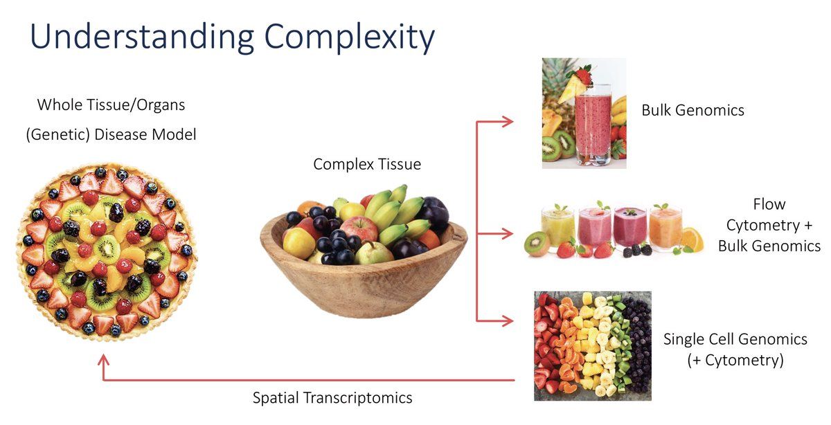
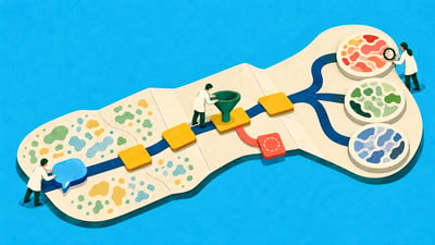
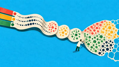
Technology has advanced from the bulk era, in which cells were pooled together to generate a single composite signal, through single-cell analysis, which provides information about individual cells, to methods that can now preserve the spatial location of each cell. (Source: LGMartelotto on X)
Suppose a tumor contains 100 immune cells. A test that analyzes all cells in aggregate might predict a favorable prognosis simply because immune cells are abundant. But when we examine the actual spatial context, we may find that those immune cells are unable to approach the cancer cells and remain confined to the surrounding tissue.
The important question is not simply how many immune cells are present, but why they cannot attack the cancer cells. The answer often lies somewhere other than the immune cells themselves.
What Spatial Biology Reveals: Immune Cells, Fibroblasts, and Endothelial Cells
The development and success of cancer immunotherapy have led many researchers to focus on immune cells within the tumor. Yet one of the most powerful insights offered by spatial biology is the role of fibroblasts as physical and biological barriers.
In the past, fibroblasts were often regarded merely as structural components supporting cancer cells. Spatial analysis, however, has revealed that they can play a major role in shaping cancer behavior.
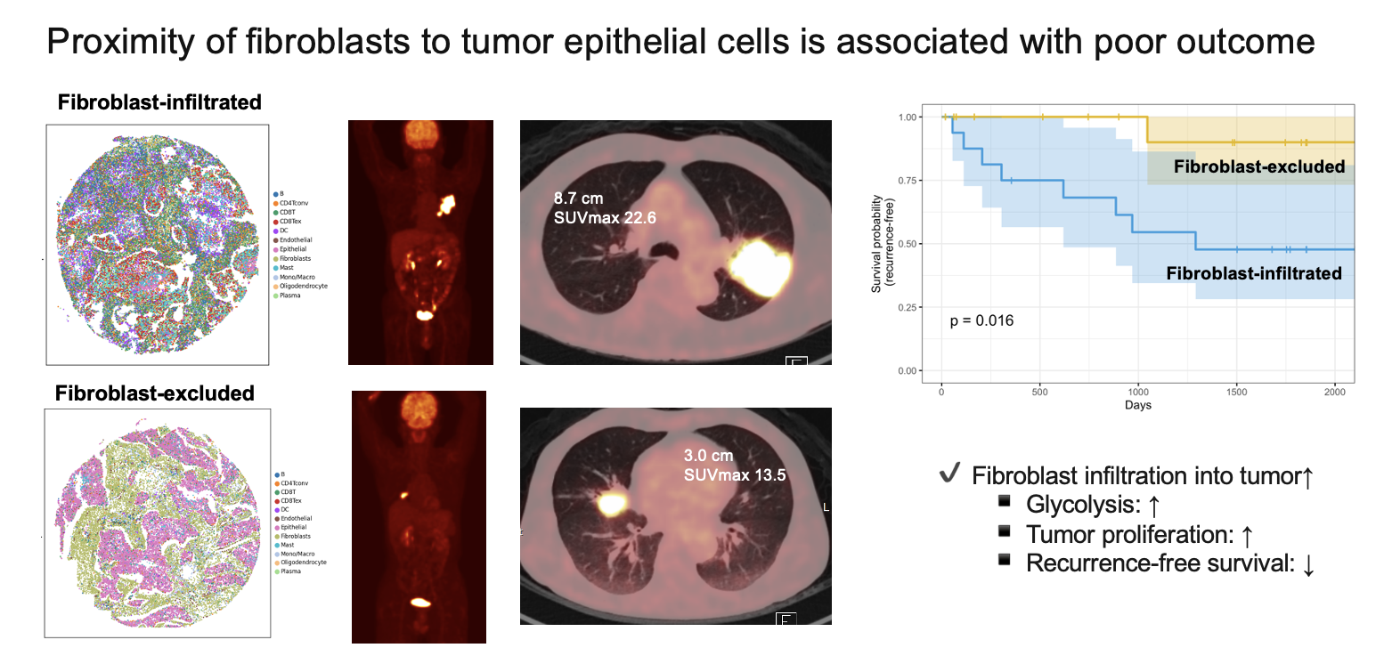
Findings from Portrai’s previous research showing that cancer aggressiveness may vary according to the degree of fibroblast infiltration and the spatial arrangement of fibroblasts relative to cancer cells. (bioRxiv, 2024: https://www.biorxiv.org/content/10.1101/2024.05.16.594592v1)
If we were to reanalyze Patient B’s tumor spatially, we might expect it to differ from Patient A’s in several respects. These differences could include the extent to which cancer-killing immune cells infiltrated the tumor, the spatial proximity between cancer cells and fibroblasts, and fibroblast barriers associated with prognosis that may prevent immune cells from reaching the cancer.
Conventional bulk or single-cell testing might report that “T cells are abundant” and therefore predict that immunotherapy will be effective. A physician who can see the spatial context, however, may reach the opposite conclusion: “T cells are abundant, but they cannot reach the cancer cells, so the treatment may not work.”
This is the explainable rationale that spatial biology can provide.
Beyond Observation: Spatial Biology for Better Treatment Decisions
Ultimately, every technology must serve the patient. Spatial biology offers a new way to evaluate cancer where conventional methods have remained limited.
Spatial biology reveals not merely the cancer’s address, or stage, but its blueprint.
Until now, we have largely treated the “average patient.” Yet the interactions among cells within a tumor differ profoundly from one patient to another and from one tumor tissue to another. Only by understanding the unique blueprint and vulnerabilities of each patient’s cancer can we move beyond a contest of probabilities and make treatment decisions with confidence grounded in data.
This is also the path Portrai is pursuing: using AI to interpret the complex spatial information hidden within H&E slides and reveal optimal treatment pathways that TNM staging alone cannot show. From discovering new therapeutics to designing treatment strategies based on predicted responses, spatial biology is opening new ways for us to understand cancer.
Looking into the spatial world beyond the microscope is where the most precise strategies for saving patients begin.