Knocking on the Same Target — Why Do the Results Differ?
Antibody-drug conjugates (ADCs) are the hottest modality in cancer treatment right now. The global ADC market surpassed $15B in 2026, with over 200 candidates competing in the pipeline. Yet the most confounding moment in ADC development is when two drugs targeting the same antigen produce entirely different clinical outcomes.
A prominent recent example involves ADCs targeting TROP2. Let us compare two TROP2-ADCs currently competing in the clinic:
ADC-A: Uses a pH-dependent hydrolyzable carbonate linker, carrying a topoisomerase I inhibitor payload with high membrane permeability
ADC-B: Uses a protease-cleavable tetrapeptide linker, carrying a different topoisomerase I inhibitor payload
Both bind TROP2. Both carry topoisomerase I inhibitor payloads. Yet their clinical results show unexpected divergence:
Lung adenocarcinoma: ADC-B achieves significantly higher intratumoral payload concentrations compared to ADC-A
Triple-negative breast cancer (TNBC): In certain subgroups, ADC-A delivers payload more efficiently than ADC-B
Same target, similar payload class. So why does this difference occur?
The conventional wisdom of ADC development — "high target expression means good efficacy" — cannot explain this phenomenon. The answer lies hidden within the space of tumor tissue.
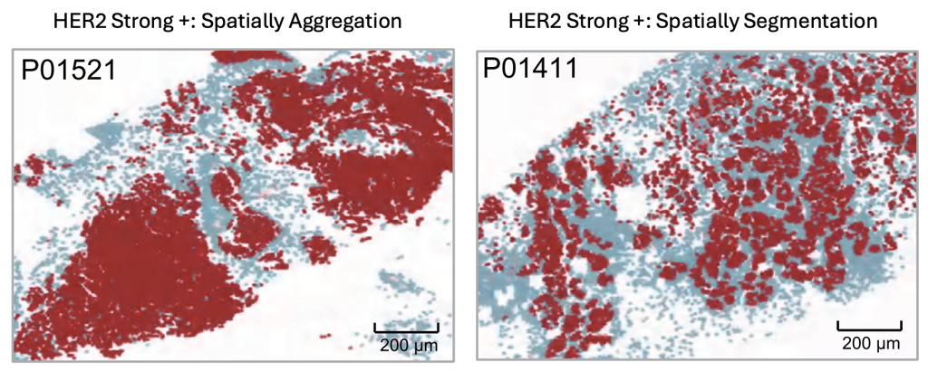
Figure 1. An example of breast cancers where HER2 is strongly positive in both cases, yet their spatial expression patterns differ. (Ma et al. Cancer Cell 2025)
The Space Where ADCs Operate: Three Hidden Variables
The factors determining ADC efficacy go beyond target "quantity" alone. From the perspective of spatial biology, several critical variables — invisible to conventional data — emerge.
1. Vessel-to-Target Distance
ADCs are administered intravenously. This means the drug must reach the tumor through vasculature. No matter how high target expression may be, if the distance from blood vessels to target-expressing cells is too great, the probability of drug arrival drops significantly. By quantifying the spatial distance between vascular endothelial cell markers (e.g., PECAM1, VWF) and target genes (e.g., TROP2, HER2) in spatial transcriptomics data, we can see that tumors with identical H-scores may have vastly different drug accessibility.
This is information that is absolutely invisible to conventional IHC or bulk RNA-seq. Grinding and analyzing the tumor yields only the conclusion "TROP2 is high," but examining the space can tell an entirely different story: "TROP2 is high, but a large proportion of high-expression zones are far from vasculature, making drug delivery difficult."
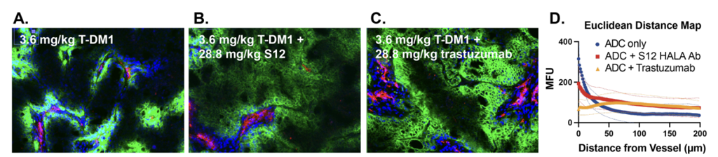
Figure 2. An experimental result showing that the concentration of antibody-class drugs (green) decreases as the distance from blood vessels (red) increases. (Kopp et al., BioRxiv 2024)
2. Spatial Distribution of Linker-Cleaving Enzymes
There is a critical element in ADC mechanism that is often overlooked: the linker must actually be cleaved within the tumor for payload to be released. Linker cleavage is carried out by specific enzymes (proteases, etc.), and whether these enzymes are expressed in the same spatial location as the target determines payload release efficiency.
ADC-A's linker is a pH-dependent hydrolyzable type, while ADC-B's linker is a protease-cleavable type. Because the release mechanisms of these two linkers differ, the spatial matching with enzyme distribution within the tumor microenvironment is crucial. Using spatial transcriptomics data, we can confirm that even among tumors with identical target expression levels, payload release efficiency can vary several-fold depending on the spatial pattern of linker–enzyme matching.
3. Spatial Range of the Bystander Effect
Another powerful weapon of ADCs is the bystander effect — the ability to kill neighboring cancer cells that do not express the target. This phenomenon, where payload released extracellularly affects adjacent cells, is particularly important in tumors with heterogeneous target expression. However, the "range" of the bystander effect varies according to the physicochemical properties of the payload (membrane permeability, half-life, etc.), and if this range is smaller than the size of target-negative clusters within the tumor, therapeutic blind spots occur.
By measuring the distance between target high-expression and low-expression regions in spatial data, we can predict whether the bystander effect can sufficiently cover a given tumor. Recent studies have reported significant correlations between these spatial distance metrics and actual clinical response.
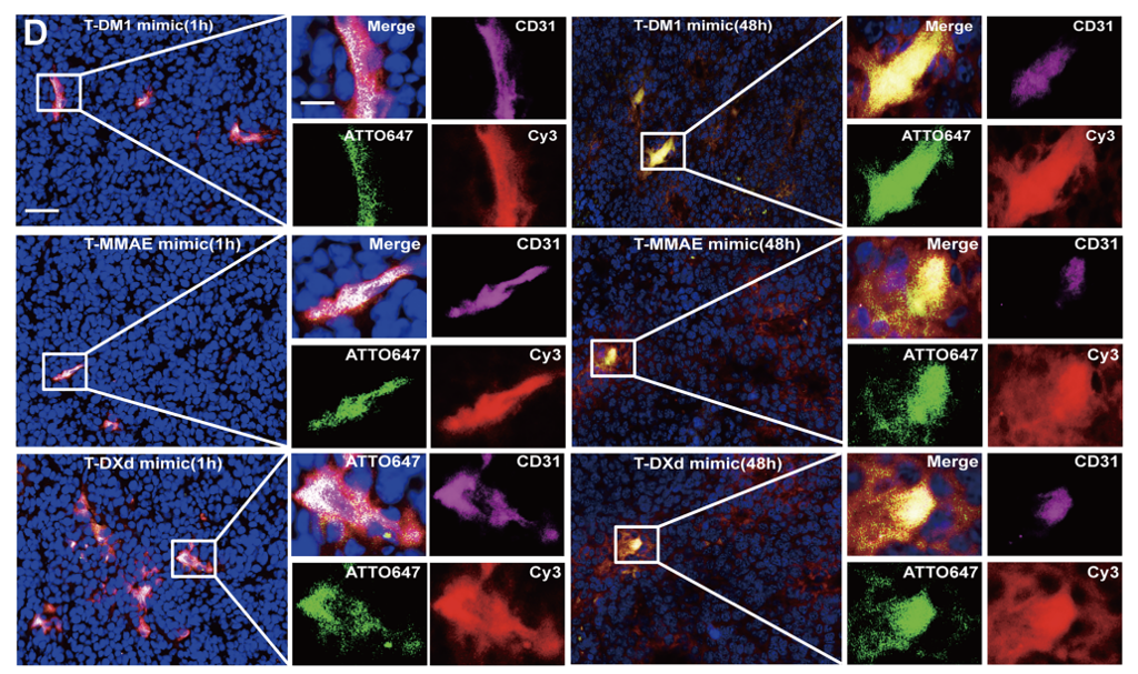
Figure 3. Bystander Effect Spatial Coverage. Compared to the distribution of antibody and vasculature (green, purple), the distribution of payload through bystander effect (red) can be seen spreading more broadly. (Wei et al., Clin Cancer Res 2024)
So, Where Does the Difference Between the Two TROP2-ADCs Come From?
When we look at these three spatial variables in an integrated manner, the clinical outcome differences between the two ADCs begin to be explained.
Why ADC-B is superior in lung adenocarcinoma:
TROP2 expression in lung adenocarcinoma is relatively concentrated in vessel-proximal regions, providing good ADC accessibility
Enzymes matching ADC-B's protease-cleavable linker are abundantly co-localized within the tumor region
As a result, payload is activated across a broader area within the tumor
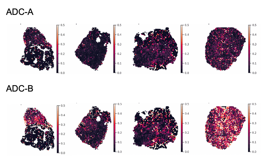
Figure 4. Portrai internal data. Results of applying the TME-PK™ model to ADC-A and ADC-B in lung adenocarcinoma, showing that significantly higher drug delivery occurs with ADC-B.
Why "Target Expression = Efficacy" Breaks Down
ADC development has long followed a relatively simple formula:
Is the target highly expressed on the tumor? → IHC H-score
Is the target low in normal tissue? → Safety margin
If both criteria are met, the target has been deemed "good for ADC." However, we now know this formula is incomplete. In last month's blog, I discussed how two numbers — "distance (20 µm) and boundary (150 µm)" — can split immunotherapy response. The same applies to ADCs. In addition to "how much is there," "where and how it is arranged" determines the drug's fate.
The questions spatial biology poses for ADC development are these:
Is the target within reachable distance from vasculature?
Are linker-cleaving enzymes in the same spatial compartment as the target?
Is the bystander effect range sufficient to cover target-negative clusters?
How do all these conditions differ by cancer type and by patient?
To answer these questions, rather than grinding the tumor to obtain averages, we must trace the drug's journey across the space of the tumor.
Simulating the Drug's Journey on Spatial Maps
It is now becoming possible to actually integrate these spatial variables and predict a drug's intratumoral behavior. On a patient's spatial transcriptomics data, the entire ADC process — binding → internalization → linker cleavage → payload release → bystander diffusion — can be simulated.
This tumor microenvironment pharmacokinetics (TME-PK™) modeling approach can address several powerful questions:
For the same target, how does changing the linker or payload alter intratumoral drug distribution?
For a specific patient's tumor, which ADC design is optimal?
For tumors where an existing ADC has failed, which parameters could be tuned for improvement?
This is no longer theoretical. By combining spatial transcriptomics data from real patient tissues with the physicochemical parameters of drugs, predicting ADC spatial behavior in silico has become reality.
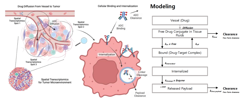
Figure 5. Portrai internal data. TME-PK™ modeling.
Conclusion: The Next Language of ADC Development Is 'Space'
ADCs are called "precision drugs." Yet ADC development to date has missed the most critical precision of all — how the drug moves within tumor tissue and where it actually works.
Spatial biology fills this gap. Not the quantity of target, but the arrangement. Not the chemistry of the linker, but where cleavage occurs. Not the potency of the payload, but the range of delivery. All of these can only be defined, measured, and optimized on spatial data.
In a $15B market where ADCs compete for the same targets, the secret behind "same target, different destiny" is no longer a black box. The answer is already written on the space of the tumor, and we have begun to read it.
References
Ma D, Dai LJ, Wu XR, et al. Spatial determinants of antibody-drug conjugate SHR-A1811 efficacy in neoadjuvant treatment for HER2-positive breast cancer. Cancer Cell. 2025;43(6):1061-1075. DOI: 10.1016/j.ccell.2025.03.017
Rubahamya B, Dong S, Thurber GM. Clinical translation of antibody drug conjugate dosing in solid tumors from preclinical mouse data. Science Advances. 2024. DOI: 10.1126/sciadv.adk1894
Kopp A, Dong S, Kwon H, et al. In vivo Auto-tuning of Antibody-Drug Conjugate Delivery for Effective Immunotherapy using High-Avidity, Low-Affinity Antibodies. BioRxiv. 2022.
dOI: 10.1102024.04.06.5884331/Wei Q, Yang T, Zhu J, et al. Spatiotemporal Quantification of HER2-targeting Antibody-Drug Conjugate Bystander Activity. Clinical Cancer Research. 2024;30(5):984-997. DOI: 10.1158/1078-0432.CCR-23-1725