
Why Most Bispecifics Stall in Solid Tumors
Bispecific antibodies are the next chapter of immuno-oncology. The design — recognizing two different antigens simultaneously, one on the tumor cell and one on the T cell (CD3) — creates a direct bridge between immune effectors and cancer.
The success in hematologic malignancies has been impressive. Blinatumomab (CD19×CD3), teclistamab (BCMA×CD3), and others have demonstrated potent efficacy.
But in solid tumors, it's a different story.
The conventional approach asks: "Are both targets co-expressed?" At Portrai, we ask: "Are both targets on the same cell, in the same space? And are they separated in normal tissue?"
This difference determines whether bispecifics succeed or fail in solid tumors.
The Co-expression Trap: The Illusion Created by Bulk Data
The first step in bispecific target pair selection typically goes like this: screen bulk RNA-seq databases for surface antigen pairs that are simultaneously highly expressed in tumors.
The problem: co-expression ≠ co-localization.
When bulk RNA-seq shows that target A and target B are both highly expressed in the same tumor, it does not mean they are on the same cell. Target A may be expressed on cancer cells in the tumor core, while target B sits on a different clone at the invasive margin. Or target A may be on cancer cells while target B is on adjacent stromal cells.
Design a bispecific against this pair, and what happens? Since both targets aren't physically on the same cell, the bispecific's dual-binding cannot engage. The drug reaches the tumor but doesn't work as intended.
Conventional approach: Bulk RNA-seq → "Both A and B are high in this tumor" → design bispecific A×B
Spatial approach: Spatial transcriptomics → "A and B co-localize on the same cell in only 32% of cases; the remaining 68% are in separate regions" → patient selection and target pair reassessment needed
Chekalin et al. (Bioinformatics Advances, 2024) addressed this problem directly. In HCC, they first selected antigen pairs with the largest tumor-versus-normal differential from bulk RNA-seq (OCTAD database), then validated true co-expression at the single-cell level using scRNA-seq (39,361 normal cells + 18,000 HCC cells). Finally, they confirmed in situ co-localization via spatial transcriptomics, identifying GPC3-MUC13 — a pair co-expressed on the surface of over 30% of malignant hepatocytes with minimal expression in other cells.
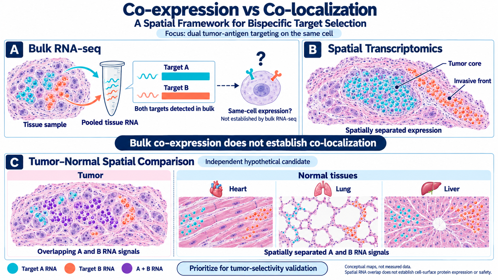
[Figure 1 — Co-expression vs. Co-localization: A Spatial Framework for Bispecific Target Selection]
Top: (A) Bulk RNA-seq shows that two targets are co-expressed, leading to the assumption that they are expressed in the same cells. (B) Spatial transcriptomics reveals their actual distribution: Target A is expressed in the tumor core, whereas Target B is expressed at the invasive front. This contrast illustrates that co-expression does not guarantee co-localization.
Bottom: (C) Place a co-localization map of the tumor (left) alongside co-localization maps of normal tissues, including the heart, lung, and liver (right). A target pair that co-localizes in the tumor but is spatially separated in normal tissues represents a safe bispecific candidate. (Schematic illustration based on the concept of the three-step pipeline described in Chekalin et al., Bioinformatics Advances, 2024.)
Tumor Side: Same Target Pair, Different Spatial Landscapes
Selecting a target pair is not the end. The same target pair shows entirely different spatial co-localization patterns across cancer types and across patients.
This is the exact same structure we discussed for ADCs in March: "Same target, different fate" — arrangement, not quantity, determines outcome.
For bispecifics, this problem is even more severe. Because bispecifics require both targets to be simultaneously accessible within binding distance to function.
Spatial Requirements for T Cell Engagers
For T cell engager-type bispecifics (e.g., CD3 × tumor antigen) to work, additional spatial conditions must be met:
Cell-level co-localization of both targets: Tumor antigen on the cancer cell surface, CD3 on the T cell surface — this is a given
Physical proximity between T cells and tumor cells: For the bispecific to "bridge" both cells, T cells and tumor cells must be within immunological synapse formation distance
Absence of immunosuppressive barriers: As discussed in our June blog, CAF barriers and immunosuppressive cell proximity can neutralize T cell function even when T cells are present
Zhang et al. (ACS Chemical Biology, 2024) quantitatively demonstrated how the geometric structure of bispecific T cell engagers determines efficacy. As spacer distance between the two binding domains increased, efficacy progressively decreased. They constructed 26 pairs of TcEs with identical sizes but different orientations, demonstrating efficacy differences driven purely by spatial geometry.
Bispecific function depends on molecular-level distance (between binding domains) as well as tissue-level distance (between T cells and tumor cells). The "physical barriers" and "proximity paradox" from June apply directly here. T cells trapped behind CAF barriers cannot form synapses with tumor cells, no matter how potent the bispecific.
Normal Tissue Side: The Spatial Definition of a "Safe Pair"
The most attractive promise of bispecifics is selectivity. By requiring dual binding, they should spare normal tissues expressing only one target.
But for this promise to hold, one prerequisite must be met: both targets must not co-localize on the same cell in normal tissues.
What conventional approaches check: "Target A expression is low in normal tissue, and target B expression is also low" (bulk RNA-seq / IHC)
What spatial approaches must verify: "Do target A and target B co-localize on the same cell in normal tissue?"
These two questions can have different answers. Even when both targets show low bulk expression in normal tissue, if they co-localize — even at low levels — on specific cell types in certain organs, on-target off-tumor toxicity can occur.
As we emphasized in our May blog when discussing ADC toxicity in normal tissues, the same level of spatial analysis applied to tumors must also be applied to normal tissues. For bispecifics, this need is even more acute, because bispecific selectivity itself is fundamentally based on tumor-specific co-localization.
The practical analyses required:
Confirm cell-type-specific co-localization rates of candidate target pairs in spatial transcriptomics of major organs — heart, lung, liver, kidney, intestine
Calculate the ratio of tumor co-localization to normal tissue co-localization
Assess whether cell types showing co-localization in normal tissue are functionally critical (e.g., cardiomyocytes, alveolar epithelial cells) → toxicity risk grading
This evaluates "Is this pair effective?" in tumor and "Is this pair safe?" in normal tissue within the same spatial framework.
Portrai's Approach: Dual-Sided Spatial Target Evaluation
Portrai addresses this problem through the following framework.
Tumor side — from spatial transcriptomics data:
Quantify cell-level co-localization rates of candidate target pairs
Analyze TME characteristics of co-localization-rich regions — T cell accessibility, CAF barrier presence, distance from immunosuppressive niches
Map cancer type-specific and patient-specific co-localization heterogeneity → predict which patients are likely to benefit from specific bispecifics
Normal tissue side — from normal organ spatial transcriptomics data:
Confirm organ-specific, cell-type-specific co-localization rates of the same target pairs
Compare tumor vs normal co-localization ratios
Evaluate functional importance of normal cells showing co-localization → toxicity risk stratification
This approach applies not only to bispecific antibodies but to any dual-targeting modality — including dual-targeting ADCs and bispecific ADCs.
Conclusion: "Good Pairs" Are Defined in Space
The theoretical appeal of bispecific antibodies is clear. But the reality in solid tumors is that target pair selection is being conducted without spatial context.
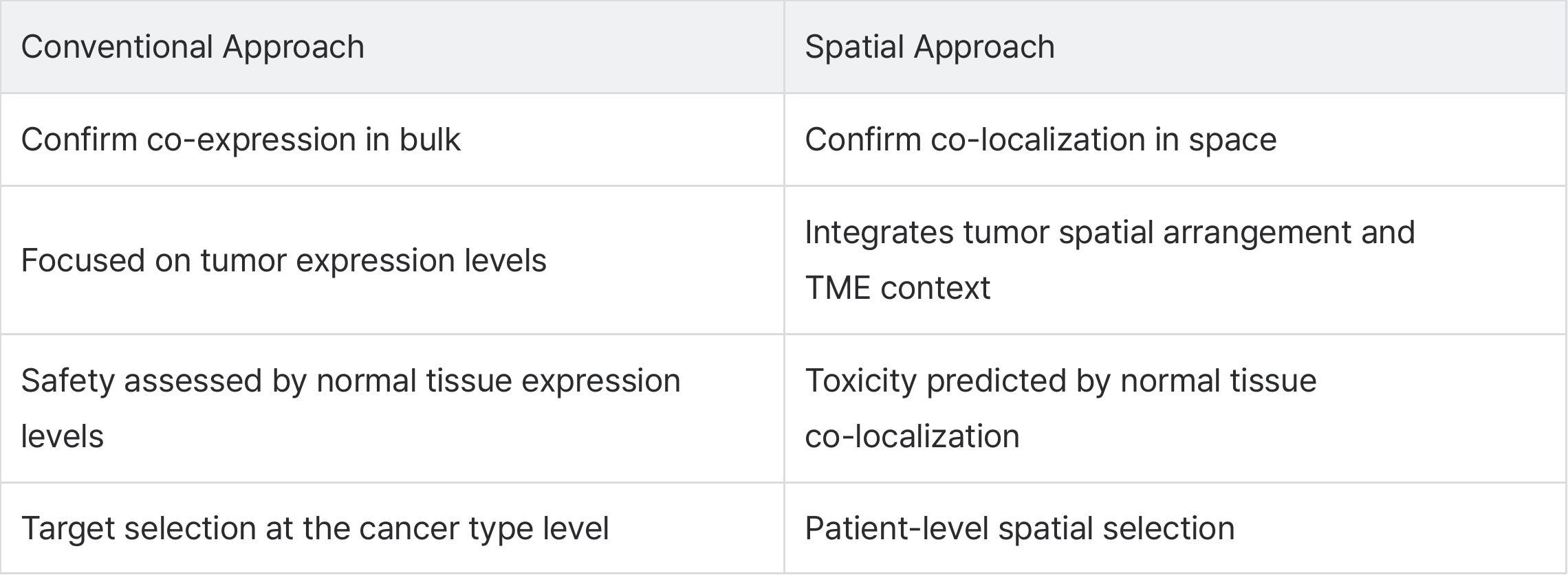
In March, we said "same target, different fate" for ADCs. For bispecifics, this extends to "same target pair, different spatial landscapes." Finding the answer among 78,000 possible surface antigen pairs is impossible with intuition or bulk data alone.
The answers are already written across the spatial landscapes of both tumor and normal tissue. Reading that space is how the promise of bispecifics becomes reality.
References
Chekalin E, Paithankar S, Shankar R, et al. Computational discovery of co-expressed antigens as dual targeting candidates for cancer therapy through bulk, single-cell, and spatial transcriptomics. Bioinformatics Advances. 2024;4(1):vbae096. DOI: 10.1093/bioadv/vbae096
Zhang Y, et al. Geometric Antibody Engineering Reveals the Spatial Factor on the Efficacy of Bispecific T Cell Engagers. ACS Chemical Biology. 2024;19(4):916-925. DOI: 10.1021/acschembio.3c00728
Nahle T, Shah V, Farraj SA, Atoui A. Next-generation T cell engagers in oncology: Pharmacologic evolution from bispecific to trispecific antibodies. Journal of Pharmacology and Experimental Therapeutics. 2025;392(12):103704. DOI: 10.1016/j.jpet.2025.103704
Portrai, AACR 2026 Poster. Spatial Transcriptomics uncovers pharmacokinetic barriers and tumor-intrinsic determinants of resistance to trastuzumab deruxtecan in breast cancer.
Portrai, AACR 2026 Poster. Spatial Transcriptomics reveals core resistance niches distinguishing non-MPR from MPR in NSCLC after neoadjuvant chemoimmunotherapy.
관련 콘텐츠